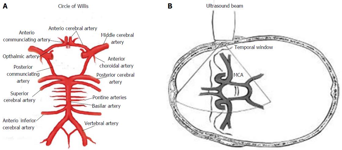

The recorded Doppler signals may be a mixture of distinct Doppler frequency shifts, which results in a spectrum showing on the TCD monitor of individual RBC’s flow due to the laminar blood flow ( DeWitt and Wechsler, 1988). The “Doppler shift frequency,” or variation in frequency between emitted and reflected waves, is directly proportional to the velocity of RBC’s circulation (BFV). Transcranial ultrasonic waves from the Doppler probe reflect off of circulating RBC’s in intracerebral vessels. TCD ultrasonography is based on the Doppler effect and the Bernoulli principle, which uses sound waves to move more quickly through the body. This review does not cover all of the concepts and interpretations of the TCD results, however, we did provide an overview. The ACA, MCA, and PCA, the ophthalmic and carotid siphons, and the vertebral and basilar arteries are among the arteries that can be investigated ( D’Andrea et al., 2016b). For the TCD scan, the ultrasound transducer is first placed on the temporal bone, over the closed eyelid, and on the base of the skull to capture the signals. In this view, TCD revolves around the Circle of Willis ( Spencer and Whisler, 1986). Moreover, vasoactive medications including drugs with vasodilatation and vasoconstriction properties results in the increase and decrease in FV ( Kassab et al., 2007).Ī thorough TCD evaluation needs to include measurements from each of the four windows, and the path of blood flow within every main branch of the circle of Willis ( Figure 2) should indeed be examined, despite the fact that each window has distinct advantages for particular arteries. While increased blood viscosity results in the elevation in FV. TCD guidelines for clinicians were published by the American Academy of Neurology which have been shown in Table 1 (Sloan et al., 2004).Īs indicated in Table 1, each vessel has a unique depth range, flow direction, and appropriate age-associated flow velocity (FV) range nevertheless, these data are influenced by a variety of physiological and pathological conditions, such as increase in age, CSF pressure, and central venous pressure lead to the decrease in the FV. In the flexed neck, the submandibular window can be used to detect the distal ICA at a 40–60 mm depth ( Alexandrov et al., 2011).

The vertebral and basilar arteries can be insonated using a flexed neck and the suboccipital window. The ophthalmic artery (OA) and carotid siphon can be evaluated through the transorbital window. The transtemporal window can also insonate the ICA bifurcation at the underlined depths with flow ( Fodale et al., 2007 Purkayastha and Sorond, 2012). When the intracranial carotid artery (ICA) bifurcation terminates in the anterior, middle, and posterior cerebral arteries (abbreviated as ACA, MCA, and PCA, accordingly), it can be identified at depths of 55–65 mm with the simultaneous flow toward or outward from the probe.
#TRANSCRANIAL DOPPLER DEPTHS WINDOWS#
The anterior, middle, and posterior windows make up the transtemporal window. Panels (B–D) show transcranial acoustic windows used in TCD evaluation, including transorbital, suboccipital, and transtemporal windows, accordingly ( Bathala et al., 2013). Panel (A) indicates ultrasonic probes, with blue arrows indicating TCD and yellow arrows indicating Transcranial color-coded sonography (TCCS). Figure 1 illustrates a variety of acoustic windows. In adults, the four most frequently used acoustic windows are temporal, suboccipital, orbital, and submandibular. It is only feasible to insonate the cerebral arteries via the acoustic window while using a low-frequency probe ( Basri et al., 2021). TCD’s ease of use as a diagnostic approach could lead to an increase in its utility in clinical and research settings for a variety of cerebrovascular conditions ( Bhogal, 2021). When dealing with cerebrovascular complications, TCD is considered to be the most practical technique to keep track of vascular alterations in response to treatment.

Such parameters provide physiologic information that can be used in combination with structural information taken from a variety of existing imaging techniques ( Purkayastha and Sorond, 2012). In the basal cerebral arteries, the TCD imaging tool uses low-frequency ultrasonic waves (i.e., ≤2 MHz) to evaluate blood flow parameters and cerebrovascular hemodynamics in real-time.


 0 kommentar(er)
